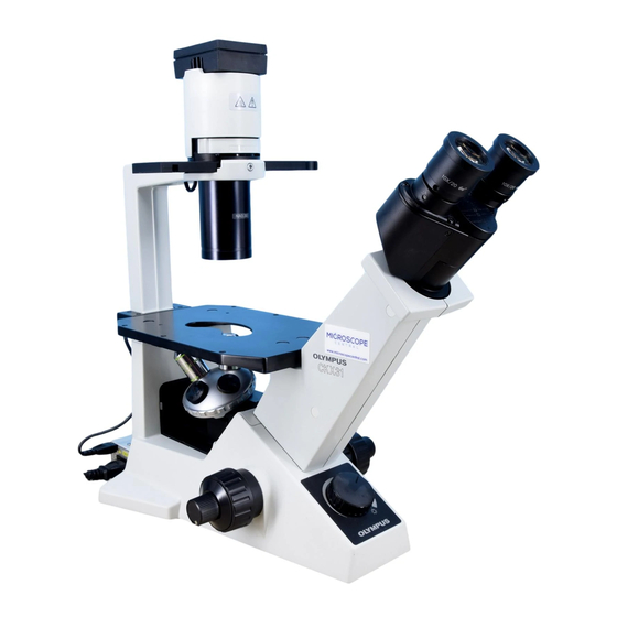
Olympus CKX31 Manual de reparación - Página 4
Navegue en línea o descargue pdf Manual de reparación para Microscopio Olympus CKX31. Olympus CKX31 43 páginas. Culture microscopes
También para Olympus CKX31: Visión general (7 páginas), Folleto y especificaciones (4 páginas)

CKX31/CKX41
1. Outline
(1) CKX31: Inverted culture microscope with fixed observation tube
(successor to CK30)
CKX41: Inverted culture microscope with interchangeable observation tube
(successor to CK40)
Reflected light fluorescence attachment mountable
(2) Target market and applications
Tissue culture routine market represented by culture, growth, immunity and pharmacy.
(3) External standards acquired
The product complies with UL. The CE mark was self-declared.
(a) IEC1010-1
(b) UL3101-1
(c) EN55011 Group 1 class B
(d) EN50082-2(1995)
(4) Service life
8 years
2. Features
CKX31/CKX41
(1) UIS optical system is adopted and it brings drastic improvement in image flatness as well as
excellent clarity right to the edge, even in the case of wide field-of-view images.
(2) Use of a phase slider makes it possible to achieve high contrast images(active cell).
1) Common phase contrast ring slit(RS) is provided for 10X-40X to enable phase contrast
observation of 10X-40X simply by changing the objective.(when combined with IX2-SLP)
2) No centering is necessary for phase contrast ring slit (RS) and phase membrane in objective.
(Applicable when the CKX31/CKX41 is combined with the IX2-SLP.)
Slider
IX2-SLP
IX2-SL
(3) Main switch and light intensity adjustment knob are located on the front of microscope frame.
(4) Specimen in various types of containers can be observed by selecting effective observation
methods.
1) Observation can be made by using various observation methods.
Observation method
Brightfield observation
Phase contrast observation
Fluorescence observation
(5) The working distance(W.D.) is 72mm with condenser and 150mm without condenser.
Therefore, various containers from petri dishes to roller bottle and squareflasks can be used
for observing specimens.
(6) Designed with the minimum necessary footprint, working space can be secured even in confined
space like a clean bench.
A. OUTLINE OF PRODUCT
10X
4X
Common to 10X - 40X (built-in)
dedicated RS
(built-in)
PHL
IX2-SLPH1
(built-in)
IX2-SLPHC
Observation of suspended cells
Observation of adhering cells
(Observation of the internal structure)
Observation of GFP appearance
Magnification
40X
20X
IX2-SLPH2
(RS adjustment:
unnecessary)
Application
A-1
RS adjustment
Unnecessary
Necessary
