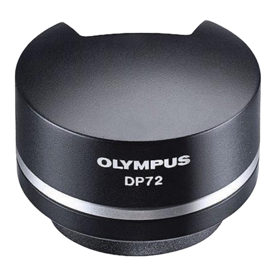
Olympus DP72 Instrukcja obsługi - Strona 6
Przeglądaj online lub pobierz pdf Instrukcja obsługi dla Aparat cyfrowy Olympus DP72. Olympus DP72 40 stron. Microscope digital camera

1
Conformity of the System
Restrictions in Use
1. The applicable camera adapter is the U-TV0.5XC-3, U-TV0.63XC, MVX-TV0.63XC or the combination U-TV1X-2 + U-CMAD3.
The U-TV0.5XC should not be used because it deteriorates the image flatness.
A camera adapter with a magnification below 0.5X cannot be used because part of image will be cut off.
2. When the DP72 is connected to the rear port of the U-DPT or U-MPH, the peripheral part of the recorded image may be
deteriorated due to the optical performance of the U-DPT or U-MPH.
3. When the U-TV0.5XC-2 or U-TV0.5XC-3 is used, using two or more intermediate attachments* may obscure or cut off the
peripheral part of the field of view or may make flare noticeable.
* Example of two intermediate attachments with BX microscope: Vertical illuminator + Intermediate attachment with a
length equivalent to the U-CA
4. Under fluorescent ring illumination or other AC-driven illumination such as a phase control light intensity adjusting
illumination system, the following phenomena may be observed when the light intensity is increased and exposure time
is decreased:
· Flickering of the displayed image.
· Instability in exposure.
· Hatching patterns in pixel shift recording (4140 x 3096 or 2070 x 1548 pixels).
However, provided that the brightness can be adjusted using the light intensity control knob or ND filters, the above
phenomena may be attenuated by adjusting the brightness so that the exposure time exceeds 1/50 sec.
For details on the microscope models using AC-driven illumination, contact Olympus.
5. Non-Olympus microscopes and commercially available C-mount lenses can be used provided that they match a CCD
with a size of no less than 2/3 inch and the lens projection length from the C-mount body attaching section is no more
than 6 mm. However, problems due to optical adaptability, such as shading, may be observed.
6. When the specimen has a low contrast (near transparent) or high reflectance (mirror status) and the aperture iris diaphragm
is stopped down near the smallest aperture, spot flare may be noticeable.
7. When the edge of a non-transmitting object is observed under the STM6 transmitted illumination, flare may be noticeable
due to the difference in brightness between the transmitted sections (over-exposure) and non-transmitting section (under-
exposure). To reduce the flare, set a lower exposure using the exposure correction function or setting the exposure
manually.
8. When a low-power objective (below 4X) is used, the peripheral part of the field of view may be obscured. In this case, use
an ultralow-magnification condenser (U-ULC-2).
9. When the U-CFU is used, it is required to set the exposure to a longer period than 1/30 sec. using the manual exposure
mode and control the brightness by engaging or disengaging ND filters.
10. Red, horizontal flare due to surface reflections of the area outside the CCD's effective image pickup area may sometimes
be observed on the upper part of the image under the following conditions.
· During brightfield observation of a specimen with a large difference in brightness, particularly when the bright part
of the specimen comes on the upper part of the image.
· When the Aperture iris diaphragm is stopped down to the minimum aperture.
11. When a specimen with high reflectivity is observed with reflected light brightfield observation through the eyepiece/
camera light path of a trinocular tube using a 0.5X camera adapter such as the U-TV0.5XC or U-TV0.5X, the image in the
area outside the CCD's effective image pickup area may b observed as vague ghosts in the peripheral area of the visual
field of eyepiece.
12. Flare may be produced during reflected light darkfield observation under overexposure To reduce the flare, use the
exposure correction function or reduce the exposure with manual exposure control.
13. During recording with image shifting (4140 x 3096 or 2070 x 1548 pixels), the image may be disturbed if the specimen is
moved.
14. If the camera or microscope is vibrated during recording of a 4140 x 3096 or 2070 x 1548 pixel image, the image will be
disturbed. Note that the factors causing vibrations include operation of the keyboard or mouse on the same desktop
where the microscope and camera are installed.
Operating Environment
Temperature: 10 to 35°C. Humidity: 20% to 85% (without condensation).
See page 29 for details.
Recommended Monitor Specifications
· A monitor with the 1280 x 1024 or larger full-color display capability.
· An Adobe RGB compatible monitor, provided that the camera head is used in the Adobe RGB mode.
3
