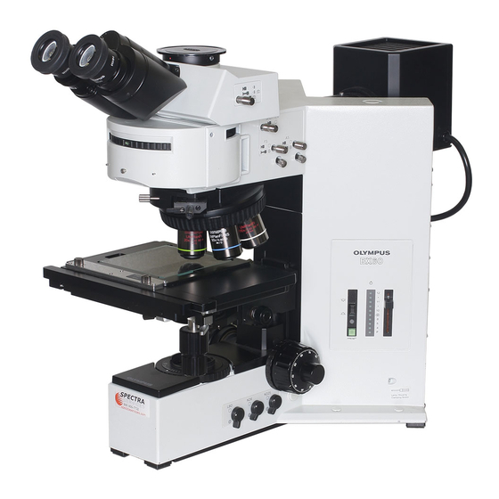
Olympus BX60 Manual de instrucciones - Página 38
Navegue en línea o descargue pdf Manual de instrucciones para Microscopio Olympus BX60. Olympus BX60 48 páginas. System microscope
También para Olympus BX60: Manual de inicio rápido (4 páginas)

Ottsefvaiflbn
1. Place the specimen on the stage and move the stage to bring the specimen into focus.
2 Adjust the field iris diaphragm until the diaphragm opening circumscribes the field of view.
3. Stopping down the aperture iris diaphragm somewhat may increase the contrast
4. Rotate the prism control knob 0 of the DIC prism slider to adjust the interference color of the background, and
tQ achieve maximum contrast depending on the specimen under observation, as outlined below:
(II Rotating the prism control knob of the slider win continuously change the interference color of the background
from gray to magenta (-100-600 nm).
• if the background color is black ((tatter fringe), darkfietd like observation is possible.
• if the background color is gray, a three-dimensional looking; image with maximum contrast with grey sensitivity
can be obtained.
• If the background color is magenta, even a minor optical retardation can be observed as a color change.
• Care should be taken to keep the specimen surface dean, as even a small amount of contamination on
the surface may show up dua to the exceptionally high sensitivity of the differential interference contrast
method.
121 As differential interference contrast exhibits directional sensitivity, the use of a rotatable stage is recommended.
SKTand DarWTeld 0
1. Loosen the DIC clamping screw 0 at tho front of the revolving nosepiece. and gentfy pun the U-OICR deferential
interference contrast prism <J> out Insert the dummy slider until a dick is heard. Tighten the damping screw again.
2. Disengage both the U-AN360 analyzer and the U-PO polarizer from the light path. Rotate the turret to disengage
the U-MDIC differential interference contrast cube.
BX60
0 To prepare for simple polarized light observation using the vertical illuminator, perform step fl and
6-3 Reflected Light Nomarski Differential Interference Contrast Observation outlined on page 33.
in Section
V Place the specimen on the stage and then operate the coarse and fine focus knobs to bring the specimen into focus.
Simple polarized fight observation is now possible.
2. Adjust the field iris diaphragm until the diaphragm opening circumscribes the field of view.
3. Stepping down the aperture iris diaphragm may increase the contrast somewhat
34
