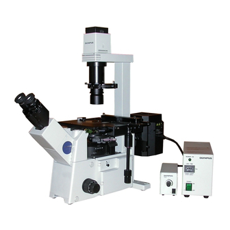
Olympus IX71 Brochure & Specs - Page 14
Parcourez en ligne ou téléchargez le pdf Brochure & Specs pour {nom_de_la_catégorie} Olympus IX71. Olympus IX71 16 pages. Microscope z-axis motor drive
Également pour Olympus IX71 : Manuel d'installation (11 pages)
FRET
Bright, simultaneous two-wavelength imaging using the primary image.
FRET Split imaging system
• Simultaneous two-color split imaging with one CCD camera.
• Unique design splits the primary image for the highest efficiency
and light transmission necessary for weak fluorescence signals
such as CFP/YFP FRET experiments.
•Compact and space-saving design takes advantage of the 70 mm
of free space between the microscope frame and the primary
image plane found on all Olympus Research Upright and Inverted
Microscopes.
• Simple cassette mechanism makes it easy to switch between
split and full frame imaging.
• Unit is up to 10% brighter than similar relay lens based, image
splitting systems.
• When used with the rectangular field stop U-RFSS, excitation
energy is limited to the camera's field of view, minimizing specimen
photo-bleaching.
Rectangular field
stop/U-RFSS
Cube cassette for
full image
Cube cassette for
Split primary image
split images
camera port/U-SIP
25
YFP
CFP
HeLa cell, in which YC3.1 (cytoplasm) and YC3.1nu (with nuclear localization signal) are coexpressed.
FRET changes are observed through histamine stimulation, and images are acquired at intervals of
50 msec.
Rectangular field stop
Specimen
U-RFSS
Objective
L shape fluorescence illuminator
Fluorescence mirror unit
IX2-RFAL
Filter set such as
XF88-2(OMEGA) or
31044v2(CHROMA)
Split primary image
Filter sliders
Built-in separation
camera port
(Emission, ND sliders)
dichromatic mirror
U-SIP
(DM505)
Excitation filter
Tube lens
Prism
High resolution camera
Research inverted
system microscope
IX81/IX71
Cube cassette for
YFP
CFP
split image
Split image
Mirrors
U-SIP main specifications
Microscope
IX71/81
Image separation
Right and left 2-separation
(can be adjusted independently)
Built-in separation dichromatic mirror
DM505 (special size)
Filter slider
Emission, ND filters' size ø25 mm,
total thickness: 8 mm
Used together with commercially available filter set
(XF88-2 OMEGA) or 31044v2(CHROMA)
Field Number
Split image: 8
Full image: 11
Magnifications
1X (primary image)
Objectives
40X and higher
Camera mounting
C-mount
Recommended camera
Chip size 2/3 inch
* Not available in some areas
Photoactivation
Photoactivation illuminator for inverted microscopy.
Photoactivation Fluorescence
Microscope system
The photoactivation illuminator allows the exposure by UV light to
specific regions of a cell for photoconversion, the uncaging of
compounds and the photoactivation of specific fluorochromes.
• A specified area of the cell can be exposed to UV light while
observing the targeted cell by fluorescence or transmitted (DIC)
method.
• Compliance with FRAP or FLIP
experiments (by special order).
• Easy system upgrade by
attaching double lamphouse
illuminator IX2-RFAW to IX2
series inverted microscope.
Double lamphouse illuminator IX2-RFAW
Setting up example for Kaede
Magnetic shutter
Double lamp
Sample
house illuminator
IX2-RFAW
Excitation filter
BP330-385
Filter slider
Pinhole slider
Field
stop
position *1
Pinhole or slit
Composed dichromatic
Filter slider
mirror of right and left path
Inverted microscope
IX series
Filter wheel 1
such as Lambda10-2
Excitation filter
475AF20(XF1072 OMEGA) or
HQ475/20x(CHROMA)
Excitation filter *2
550DF30(XF1021 OMEGA)
75W xenon apo
lamp housing
U-LH75XEAPO
Power supply unit
The novel Kaede gene is useful in biology because it exhibits photoconversion. Normally, the Kaede
gene shows green fluorescence but after exposure to UV light will exhibit red fluorescence. By using
UV light to only a specific region within a labeled cell and then noting the movement of red beyond
that region, observations of internal cellular dynamics can easily be made. The photo on the left
shows a nerve cell (from a rat hippocampus) pre-labeled with green Kaede gene.
The photo on the right side was taken after the right-most cell body was exposed to a 10 µm
diameter spot of UV light for 60 seconds, thus changing the Kaede gene from green to red. Note the
translocation of the red shifted gene outside of the 10 µm spot thus indicating intracellular transport
mechanisms.
Objective
IX2-RFAW specifications
UPLFLN40XO
Microscope
Fluorescence mirror unit
Pinhole slider
U-MF2+dichromatic mirror
(DM400 on the illustration)
Exposed area on
Fluorescence filter *2
the specimen
575ALP (XF3089 OMEGA) or
Filter slider
HQ575LP(CHROMA)
Excitation filter slider
Fluorescence filter
530DF30(XF3107 OMEGA) or
Filter size
D530/30x(CHROMA)
High resolution
Composed dichromatic mirror
camera
of right and left light path
Power consumption
Dimensions
Filter wheel 2
such as Lambda10-2
*1 Field stop position is the same
position as the focus point of the
sample.
*2 Use 550DF30 excitation filter in the
filter wheel 1 and 575ALP
fluorescence filter in the filter wheel 2
when observing red Kaede protein.
Exchange of the fluorescence mirror
unit is not required.
Application System
IX81/71/51, IX70/50
2-step exchange (pinhole or slit/vacant hole)
Pinhole and slit are available on the market
(ø16 mm Melles Griot Inc. products)
Pinhole diameter
objective magnification
3-step exchange (shutter/filter pocket/vacant hole)
BP330-385 excitation filter equipped
5-step exchange (4-step filter pocket/vacant hole)
Excitation filter: ø25 mm,
thickness: 6 mm and below
ND filter: ø32 mm,
thickness: 1 mm and below
DM400 (standard)
Slide IN/OUT type
7.4 A
Width: 710 mm
Depth: 740 mm (from the front of tilting tube to
the end of the illuminator)
* Not available in some areas
26