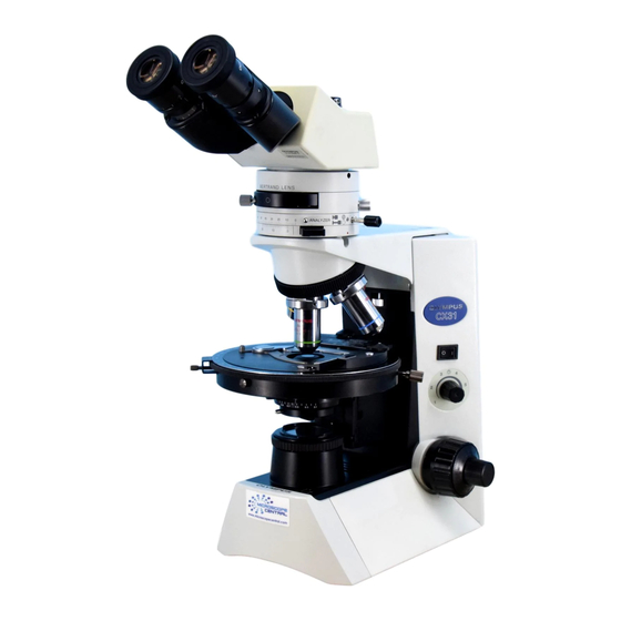
Olympus CX31-P Petunjuk Manual - Halaman 22
Jelajahi secara online atau unduh pdf Petunjuk Manual untuk Mikroskop Olympus CX31-P. Olympus CX31-P 36 halaman. Polarizing microscope

5-2 Orthoscopic Observation
}Use an objective between 4x to 100x.
1. When the U-PA intermediate tube for polarizing observation is used, set the Bertrand's lens engaging/focusing knob to the
( (OUT) position to disengage the Bertrand's lens from the light path.
2. Engage the analyzer in the light path and start observation. (Cross-Nicol position)
If open-Nicol condition is required, disengage the analyzer from the light path.
If para-Nicol condition is required, set the analyzer to the 90° position.
3. Rotate the stage to set the observation target position of the specimen dark (off position) and then rotate the stage by 45°
from there to set to the diagonal position. The retardation (R) value should be measured in this position.
4. The test plate (U-TP137 quarter-wave plate, U-TP530 sensitive tint plate) is used to produce sensitive colors and inserted in
the test plate slot. Push the plate all the way into the slot to engage the plate in the light path, and pull it till the click-stop
position to disengage it from the light path.
For the usage of other compensators, refer to their instruction manuals.
When the U-CWE quartz wedge is used, use the U-CWE2 that has the U-PA indication on the retardation reference
indication 1.
5-3 Conoscopic Observation
18
Fig. 28
}Use an objective between 20x to 100x.
1. Engage the analyzer in the light path and set to the cross-Nicol position.
2. When the U-PA intermediate tube for polarizing observation is used, set
the Bertrand's lens engaging/focusing knob to the ) (IN) position to
engage the Bertrand's lens in the light path.
3. Engage an objective between 20x to 100x in the light path.
4. Open the aperture iris diaphragm.
5. Turn the Bertrand's engaging/focusing knob to focus on the conoscopic
image as accurately as possible.
}When the U-KPA or U-OPA intermediate tube is used, remove an eye-
piece from the sleeve and look through it to view the conoscopic image.
}The image contrast may be improved by placing the interference filter
(45IF546) on the filter holder on the base of the microscope.
}If the peripheral part of the conoscopic image is dark, move the con-
denser up and down to find the height at which the peripheral part is
brightest.
1
