ATS 540 Series Manuale di istruzioni - Pagina 6
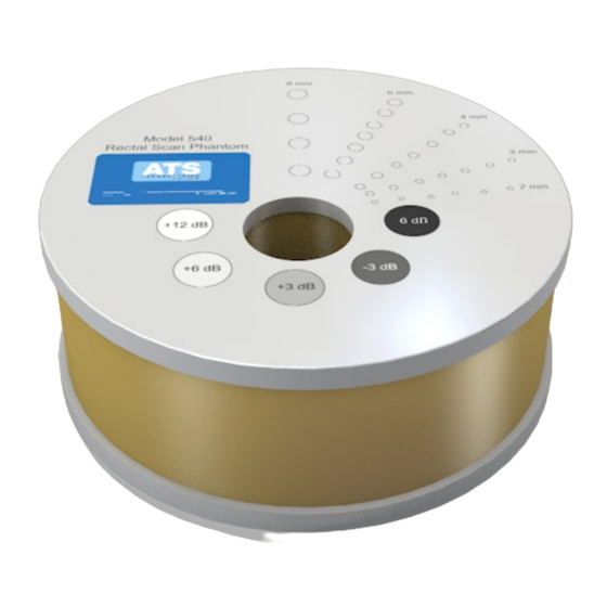
Sfoglia online o scarica il pdf Manuale di istruzioni per Apparecchiature di laboratorio ATS 540 Series. ATS 540 Series 9. Rectal scan phantom

5. Note: The test should also be performed with the output set at extreme high and low
values, in addition to the values given above. This enables any changes in output to be
more easily detected.
6. Move the transducer slightly until the smallest group of anechoic targets are clearly
displayed.
7. Freeze image and obtain a hard copy.
8. Examine the image to determine the last or deepest target structure displayed. Using
the electronic calipers or the timing markers measure the depth of this target.
9. Document the depth measurement on the quality assurance record.
Results
The depth of penetration should not shift by more than 1.0 cm, when using the same
phantom, instrument settings and transducer. If a discrepancy occurs corrective action
should be considered by the individual Ultrasound Department.
FUNCTIONAL RESOLUTION, DEFINITION AND FILL-IN
Description and Reason For Testing
Functional resolution is an imaging system's ability to detect and display the size,
shape, and depth of an anechoic structure with the test phantom. In practice, the data
obtained will give a direct indication of the minimum size structure the system is capable
of resolving at a given depth.
Definition and Fill-in describes the ability of an imaging system to detect and display the
shape and echogenic characteristics of a structure. Clinically, a correct diagnosis is
dependent upon an imaging system's ability to differentiate between a cystic or solid
structure versus echo patterns originating from the surrounding normal tissue.
Test Procedure
1. Place the phantom on a clean, flat surface with the internal scanning well positioned
for use.
2. Fill the scanning well slowly with water to avoid introducing air bubbles.
3. Insert the transducer into the scanning well.
4. Adjust the instrument settings (TGC, output, etc.) to establish baseline values for
"normal" liver scanning. If the bottom of the phantom is seen, adjust the gain settings
until the image fades and goes entirely black at the bottom of the display. Record these
settings on the quality assurance record. These setting should be used for subsequent
testing.
