Olympus VANOX Manuale di istruzioni - Pagina 25
Sfoglia online o scarica il pdf Manuale di istruzioni per Microscope Olympus VANOX. Olympus VANOX 28. Universal research microscope
I.
Use
of Immersion
Optical
Components
1. I mrnersion objectives.
1 )
Focus on the specimen
with a low-power objective.
2) Put
a drop of immersion oil
on
both the specimen and the objective front
lens.
3)
Turn the revolving nosepiece
to
bring the immersion objective into the light
path, and focus with the fine adjustment knob.
2. l immersion condensers:
1
)
Remove
t h e specimen from t h e mechanical
stage
and place a
drop
of immersion
oil on the front lens of the condenser.
2 ) Place the
specimen on the mechanical stage and slowly raise the condenser until
firm contact with the underside of
t h e specimen
slide is made.
Care
should
be
taken
to
prevent oil
bubbles from forming in the oil film between condenser
and specimen slide.
3. After use:
Carefully wipe off the immersion oil deposited on the lens surfaces with gauze
moistened with xy
lene.
Never
leave oil
on the lens surfaces after use as
oil remnants will
seriously irnpair
the performance
of
t h e
lens systems.
VIII.
Photomicrography
The Olympus
Rhotomicrographic Equipment Model PM-10 is unique1
y
qualified to be used
with
the Research
Microscope "VANOX" for routine and advanced photomicrography,
A separate, detailed instruction manual is available for the PM-10 camera system.
For
quick reference, however, you may want t o refer
to
the following poinlers when using the
PM-10.
1. Photographic
t y e p ~ e c e
Use only F K photo eyepieces for photornicro-
graphy
-
They are especially computed for this very
purpose. Insert the eyepiece into the
eyepiece
tube of
the photo
tube.
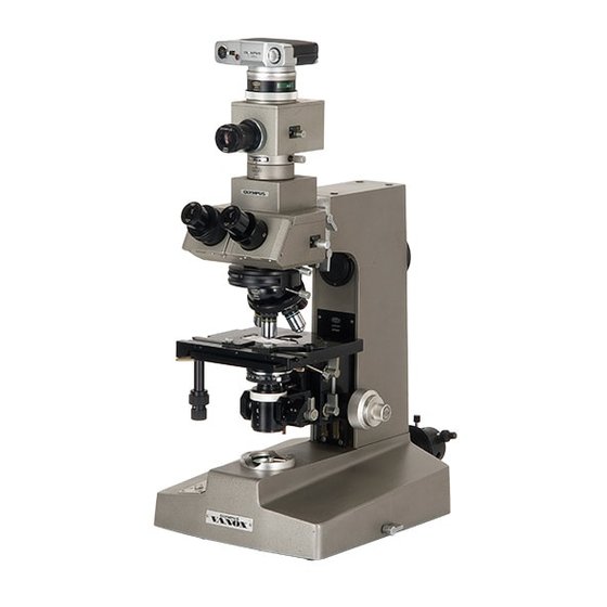
2.
Mounting the Photographic Unit
Slip
zhe
body
of
the photographic unit over the
g-
eyepiece tube
of
the photo tube. Align the dots
on photo tube and the PM-10 unit and clamp the
camera unit to the photo tube. (Fig. 20)
"When changing the photographic eyepiece, remove
as
the camera unit, exchange the
eyepiece
and
Fig. 20
re-mou n
t
the
camera
and then
replace.
3.
Setting
the Light
Path
Selector
Refer
to section H. 1
.,
page 22,
When photomicrography
is
performed on a
constant
basis
it
is recommended
to
keep the light path selector in position "CV"
(camera/viewing) and to use position
"V"
(viewing) only for the observation of weakly illuminated specimens (fluorescence,
polarization, for example).
In this case and for work with short
shutter
s p e d s ,
the
path selector is moved to position "C" (camera) for photography.