COOK Medical Extraction Balloon Gebruiksaanwijzing
Blader online of download pdf Gebruiksaanwijzing voor {categorie_naam} COOK Medical Extraction Balloon. COOK Medical Extraction Balloon 12 pagina's.
EN
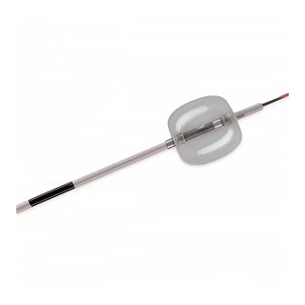
Extraction Balloon
CS
Extrakční balónek
Udtrækningsballon
DA
NL
Extractieballon
FR
Ballonnet d'extraction
Extraktionsballon
DE
EL
Μπαλόνι εξαγωγής
HU
Kiszedő ballon
IT
Palloncino estrattore
NO
Uttrekkingsballong
PL
Balon ekstrakcyjny
PT
Balão de extracção
Balón de extracción
ES
SV
Extraktionsballong
ENGLISH
INTENDED USE
This device is used for endoscopic removal of stones in the biliary system and for
contrast injection.
NOTES
This device is designed for single use only. Attempts to reprocess, resterilize, and/or reuse may
lead to device failure and/or transmission of disease.
If the package is opened or damaged when received, do not use. Visually inspect with particular
attention to kinks, bends and breaks. If an abnormality is detected that would prohibit proper
working condition, do not use. Notify Cook for return authorization.
Do not use this device for any purpose other than the stated intended use.
Store in a dry location, away from temperature extremes.
Use of this device restricted to a trained healthcare professional.
CONTRAINDICATIONS
Contraindications include those specific to ERCP and any procedures to be performed in
conjunction with balloon stone extraction.
Use of this natural latex rubber balloon is contraindicated in patients with a known
hypersensitivity to latex.
POTENTIAL COMPLICATIONS
Potential complications associated with ERCP include, but are not limited to: pancreatitis,
cholangitis, aspiration, perforation, hemorrhage, infection, sepsis, allergic reaction to contrast
or medication, hypotension, respiratory depression or arrest, cardiac arrhythmia or arrest.
Additional complications that can occur during endoscopic balloon extraction include, but are
not limited to: stone impaction, localized inflammation, pressure necrosis.
PRECAUTIONS
Refer to package label for minimum channel size required for this device.
The coordination of endoscope channel size with compatible devices is essential in obtaining
optimal results during a procedure. Refer to product package label for the minimum channel
size required for this device.
The wire guide diameter and the inner lumen of the wire-guided device must be compatible.
Assessment of the size of the stone and ampullary orifice must be made to determine the
necessity of sphincterotomy. In the event sphincterotomy is required, all necessary precautions
must be followed.
Injection of contrast during ERCP must be monitored fluoroscopically. Overfilling of the
pancreatic duct may cause pancreatitis.
SYSTEM PREPARATION
Verify balloon integrity prior to use by attaching the enclosed syringe to stopcock and inflating
balloon with air only. If any leakage is detected, do not use. Please notify Cook for return
authorization.
Flush injection port with contrast to expel all air.
Note: The stopcock is in the open position, allowing access to the balloon, when the two arms
are aligned with the catheter and syringe. To maintain balloon inflation, turn the stopcock
arm 90°.
INSTRUCTIONS FOR USE
I.
If using Fusion Quattro Balloon and a prepositioned short wire. (See fig. 1)
1.
Unlock short wire from Wire Guide Locking Device and advance tip of balloon catheter onto
prepositioned wire guide, ensuring wire guide exits catheter at Zip port.
2.
Introduce deflated balloon into endoscope accessory channel and relock wire guide. Then
advance device in short increments until it is endoscopically visualized exiting endoscope.
3.
Using fluoroscopic monitoring, position deflated balloon above stone to be removed.
Note: If more than one stone is to be removed, extract one stone at a time.
After verifying desired position of balloon, inflate balloon with air only. Note for multiple
4.
sizing balloons: Inflate balloon using fluoroscopic monitoring until balloon is visualized
occluding duct. If desired, adjust size of balloon by using reference marks on syringe.
To achieve smallest balloon size, inflate balloon to next largest size and gently pull back
on syringe to initial size. Lock stopcock.
5.
Using fluoroscopic visualization and keeping endoscope elevator open, gently withdraw the
inflated balloon toward the papilla. Warning: Do not exert excessive pressure on ampulla
while extracting stones. If stone does not pass easily, reassess need for sphincterotomy.
6.
Once balloon is endoscopically visualized in duodenum, turn stopcock to open position and
deflate balloon.
7.
Repeat extraction process, one stone at a time, until duct is clear.
Note: The previously placed wire guide may be left in position, in order to facilitate introduction
of other wire-guided devices.
If wire guide is to remain in place while device is withdrawn, utilize following steps:
8.
Remove device from endoscope accessory channel until resistance is achieved. Unlock wire
guide from Wire Guide Locking Device and perform a short segment wire guide exchange.
Relock wire guide into Wire Guide Locking Device and remove device from endoscope
accessory channel.
II.
If using Fusion Extraction Balloon and a prepositioned short wire. (See fig. 2)
1.
Unlock short wire from Wire Guide Locking Device and advance tip of balloon catheter onto
prepositioned wire guide ensuring wire guide exits catheter at IDE port.
REFER TO STEPS 2-7 IN "SECTION I" , THEN RESUME WITH STEP 2 BELOW:
Note: Previously placed wire guide may be left in position, in order to facilitate
introduction of other wire-guided devices.
2.
Prior to removing device, utilize reference marks on catheter to ensure IDE port is within
ductal system.
3.
Fluoroscopically visualize radiopaque band at IDE port. Retract wire guide until radiopaque
distal tip of wire guide passes band; a disengagement from wire guide lumen will occur.
4.
Advance disengaged wire guide to maintain ductal access.
5.
Lock wire guide into Wire Guide Locking Device and remove device from endoscope
accessory channel.
III. If using Proximal Wire Port (PWP) and a prepositioned long wire guide. (See fig. 1)
Note: For best results, wire guide should be kept wet.
1.
Remove stylet.
2.
Advance tip of balloon catheter over prepositioned long wire guide, ensuring wire guide
exits catheter at PWP.
3.
Advance deflated balloon in short increments through accessory channel using standard
long wire exchange technique until it is visualized exiting endoscope.
REFER TO STEPS 3-7 IN "SECTION I" , THEN RESUME WITH STEP 4 BELOW:
4.
Remove balloon using standard long wire exchange technique.
Upon completion of procedure, dispose of device per institutional guidelines for
biohazardous medical waste.
ČESKY
URČENÉ POUŽITÍ
Toto zařízení je určeno k endoskopickému odstraňování kamenů v biliárním traktu a k injekci
kontrastní látky.
POZNÁMKY
Toto zařízení je určeno pouze k jednorázovému použití. Pokusy o opakované ošetření
prostředku, jeho resterilizaci a/nebo opakované použití mohou vést k selhání prostředku
a/nebo k přenosu onemocnění.
Pokud je obal při převzetí otevřen nebo poškozen, zařízení nepoužívejte. Proveďte vizuální
kontrolu zařízení; věnujte přitom pozornost zejména zauzlení, ohybům a prasklinám. Pokud
objevíte anomálii, která by bránila správné funkci, zařízení nepoužívejte. Požádejte společnost
Cook o autorizaci pro vrácení zařízení.
Nepoužívejte toto zařízení pro žádný jiný účel, než pro který je určeno.
Skladujte na suchém místě, chraňte před extrémními teplotami.
Tento přístroj smí používat pouze vyškolený zdravotnický pracovník.
KONTRAINDIKACE
Ke kontraindikacím patří kontraindikace specifické pro endoskopickou retrográdní
cholangiopankreatografii (ERCP) a pro veškeré postupy prováděné v souvislosti s balónkovu
extrakcí kamenů.
Použití tohoto balónku z přírodního latexu je kontraindikováno u pacientů se známou
přecitlivělostí na latex.
POTENCIÁLNÍ KOMPLIKACE
Potenciální komplikace spojené s endoskopickou retrográdní cholangiopankreatografií (ERCP)
zahrnují, kromě jiného, následující: pankreatitidu, cholangitidu, aspiraci, perforaci, krvácení,
infekci, sepsi, alergickou reakci na kontrastní látku nebo lék, hypotenzi, ztížené dýchání nebo
zástavu dýchání, srdeční arytmii nebo srdeční zástavu.
Mezi další komplikace, ke kterým může dojít v souvislosti s endoskopickou balónkou extrakcí,
mimo jiné patří: okluze kamenem, lokalizovaný zánět a tlaková nekróza.
UPOZORNĚNÍ
Informace o minimální velikosti přístupového kanálu, potřebné pro toto zařízení, najdete na
štítku na obalu.
K dosažení optimálních výsledků během výkonu je nutná koordinace velikosti kanálu endoskopu
a velikosti kompatibilních prostředků. Informace o minimální velikosti kanálu, potřebné pro toto
zařízení, najdete na štítku na obalu.
Průměr vodicího drátu a vnitřního lumen zařízení používajícího vodicí drát musí být kompatibilní.
Pro zvážení nutnosti sfinkterotomie je potřeba zhodnotit velikost kamene a ústí ampuly. Pokud je
nutné provést sfinkterotomii, musí být dodržena všechna nezbytná bezpečnostní opatření.
Injekci kontrastní látky v průběhu endoskopické retrográdní cholangiopankreatografie (ERCP)
je nutné monitorovat skiaskopicky. Přeplnění pankreatického vývodu může způsobit zánět
slinivky břišní.
M E D I C A L
*18562/1013*