ATS 540 Series Instrukcja obsługi - Strona 2
Przeglądaj online lub pobierz pdf Instrukcja obsługi dla Sprzęt testowy ATS 540 Series. ATS 540 Series 9 stron. Rectal scan phantom

INTRODUCTION
Quality assurance tissue-mimicking phantoms are used to evaluate the accuracy and performance of ultrasound
imaging systems. The phantoms mimic the acoustic properties of human tissue and provide test structures within
the simulated environment. They are essential to detect the performance changes that occur through normal
aging and deterioration of system components. Routine equipment performance monitoring can reduce the
number of repeat examinations, the duration of examinations and maintenance time.
This phantom is constructed of a rubber-based tissue-mimicking material developed by ATS Laboratories. This
material extends the useful life of the phantom by avoiding problems due to melting, freezing, dehydration and
breakage from dropping which are common with hydrogel (water-based) phantoms. By eliminating these
problems, the durability, quality and reliability of this product is guaranteed for three years.
Most diagnostic imaging systems are calibrated for a sound velocity of 1,540 meters per second (mps), which is
the assumed average velocity of sound through human soft tissue. The rubber-based tissue-mimicking material
has a sound velocity of 1450 and 1473 mps at 0.5db/cm/Mhz and 0.7db/cm/Mhz respectively at room temperature
(23°C).
The anechoic target structures have been physically positioned to compensate for the differences in the speed of
sound, assuring accuracy of measurements. In those cases, in which the physical positioning of the targets has
not been "positioned-compensated," a simple measurement conversion calculation has been provided. This
calculation should be used when indicated in the test procedure.
PRODUCT DESCRIPTION
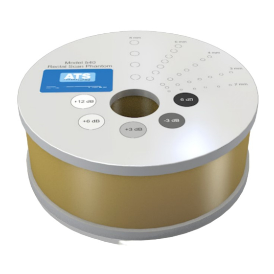
The Model #540 is designed to evaluate the performance of an imaging system's end-fired or bi-planar transrectal
rotary probes. The phantom has an internal scanning cavity to permit the insertion of a rotary probe into the body
of the phantom. The diameter of the scanning cavity is 3.5 cm (other sizes can be ordered upon request) at the
top. The cavity is tapered to prevent artifacts due to reverberation. This feature permits a 360° image to be
displayed.
The gray scale and anechoic target structures are positioned radially from the center of the scanning cavity. The
center of the first anechoic target in each row is located 1.0 cm from the edge of the scanning wall. Subsequent
targets are spaced 1.0 cm center to center, except for the 8.0 mm targets, which are spaced at 2.0 cm intervals.
The center of the scanning cavity is eccentric to the center of the phantom, as a result there are different numbers
of targets in each row.
TESTS PERFORMED
Focal Zone
•
Sensitivity
•
Functional Resolution
•
Definition and Fill-in
•
Gray Scale
•
Displayed Dynamic Range
•
