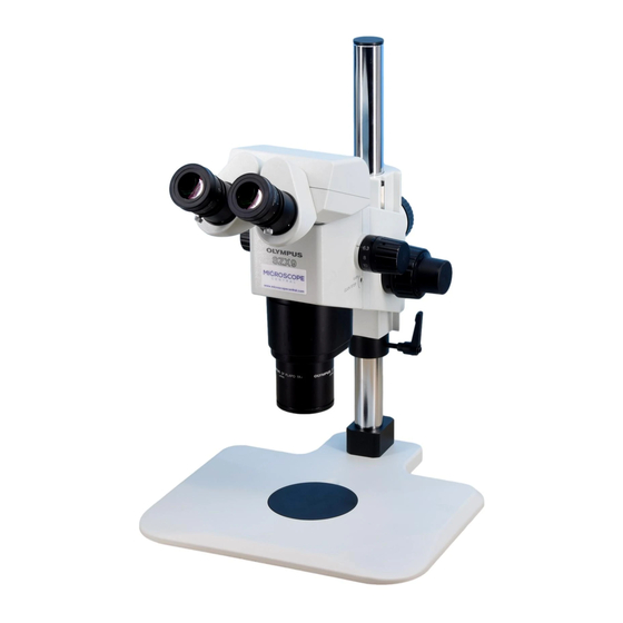
Olympus SZX-AS Instrukcja obsługi - Strona 15
Przeglądaj online lub pobierz pdf Instrukcja obsługi dla Mikroskop Olympus SZX-AS. Olympus SZX-AS 34 stron. Research stereomicroscope system

5-3 Observation Tube
@
²
@
11
Fig. 20
³
Fig. 21
1
Interpupillary Distance Adjustment
# Be sure to hold the binocular assembly @ with both hands to make
this adjustment.
While looking through the eyepieces, hold the left and right of the bin-
ocular assembly @ and adjust the eyepieces by opening or closing
them for binocular vision until the left and right fields of view coincide
completely.
Diopter Adjustment
2
(Zoom Parfocal Adjustment)
# Ensure that the eyepiece clamping knob @ is tightened.
When not using the CROSS eyepiece
1. Rotate the diopter adjustment rings ² of the eyepiece so that both scales
indicate "0".
2. Place an easy-to-observe specimen on the stage plate.
3. Rotate the zooming knob ³ to a low magnification and focus the speci-
men using the coarse and fine focus adjustment knobs.
4. Rotate the zooming knob ³ to the highest magnification and focus the
specimen using the coarse and fine focus adjustment knobs.
5. Rotate the zooming knob ³ to the lowest magnification, then focus the
specimen by rotating the left and right diopter adjustment rings instead of
the coarse and fine focus adjustment knobs.
When using the CROSS eyepiece
1. Look into the CROSS eyepiece and focus the cross lines by rotating the
diopter adjustment rings.
2. Place an easy-to-observe specimen on the stage plate.
3. Rotate the zooming knob ³ to a low magnification and focus the speci-
men looking into the CROSS eyepiece and using the coarse and fine
focus adjustment knobs.
4. Rotate the zooming knob ³ to the highest magnification and focus the
specimen using the coarse and fine focus adjustment knobs.
5. Rotate the zooming knob ³ to the lowest magnification, then focus the
specimen by rotating only the diopter adjustment ring of the non-CROSS
side of the eyepiece instead of the coarse and fine focus adjustment
knobs.
}Note (or memorize) the diopter readings of the left and right eyepiece
scales so that they can be duplicated quickly in the next observation.
(Fig. 20)
(Fig. 21)
