Olympus CKX41 Instrukcja naprawy - Strona 9
Przeglądaj online lub pobierz pdf Instrukcja naprawy dla Sprzęt laboratoryjny Olympus CKX41. Olympus CKX41 43 stron. Reflected fluorescence system
Również dla Olympus CKX41: Przegląd (7 strony), Broszura i specyfikacje (4 strony), Instrukcja obsługi (36 strony)
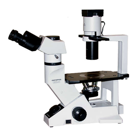
CKX31/CKX41
1. Inspection items and Methods
<CKX31>
Part
Binocular
Interpupillary
distance adjustment
tube
range
Interpupillary
distance working force
Revoving axis
Left / right optical axis On image surface:
Absolute optical axis
(center of standard
objective)
Exit pupil center
Pafocality
* 1) Since the binocular section of CKX31 is the same as that of CK30, the procedure of optical axis adjustment is
performed in the same manner as CK30. (Refer to the adjustment part of D-3 - D-14 in CK30/40 repair manual.)
2) Nesessary jigs are partially different because UIS optical system is adopted in CKX. (Refer to the above description.)
3) Diffrence between CK30 and CKX31 in parfocality adjustment: CK30 uses the spacer (selection : 3pcs. available),
While CKX31 uses the washer ( selection: 6 pcs. available) when adjusted and it requires more precise standard.
B. INSPECTION STANDARD
Item
Standard
48mm - 75mm
5N - 15N
(500g - 1500gf)
0.1mm or less on the image
surface
0.2mm or less in vertical
direction
0.2mm or less in outward
direction
0.4mm or less in inward
direction
0.1mm or less on the image
surface
Within 30% of objective's
exit pupil diameter
+ / - 0.3mm
In the observation state, insert a thin sheet of a
paper with graduations at the eye point position and
measure the interpupillary distance.
Put a string on the sleeve and measure the working
force to actuate the interpupillary distance using
a tension gauge.
Tension gauge (30N): OT3223
Using the standard eyepiece (KN0048; with
adapter-1) and an objective (PlanC10X),
observe a specimen whose center can be identified
(ex. concentric circles) on the stage.
By moving the stage, match the specimen center
and the visual field center in the right sleeve.
Open and close the interpupillary distance, and
read the movement of the image using the reticle
scale (1 graduation = 0.1mm) of KN0048.
Using the standard eyepiece (KN0048; with
adapter-1) and an objective (PlanC10X),
observe a specimen whose center can be identified
(ex. concentric circles) on the stage.
Taking the right sleeve as the reference, read the
displacement between the specimen center and
the visual field center in the left sleeve using
the reticle scale (1 graduation = 0.1mm) of the
KN0048.
Combining the standard eyepiece (KN0048; with
Adapter-1), a microscope frame (product), the
standard objective (KN0041), read the
displacement between specimen center in KN0041
and the visual field center in the right sleeve
using the reticle scale (1 graduation = 0.1mm) of
the KN0048.
Combining the centering telescope (KN0029),
an objective (PlanC10X), and microscope frame
(product), read the displacement between the exit
pupil center of the objective and cross hairs center
of centering telescope (KN0029).
Combine the standard eyepiece (KN0048; with
adapter-1, the focusing telescope (FT-36),
the microscope frame (Product), the standard
objective (KN0041).
In the left sleeve, focus on the specimen in KN0041
and check the parfocality using the helicoid scale
(1 graduation = 0.1mm) of the KN0048.
B-1
Method