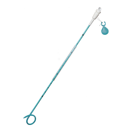
Argon Medical Devices SKATER Skrócona instrukcja obsługi
Przeglądaj online lub pobierz pdf Skrócona instrukcja obsługi dla Sprzęt medyczny Argon Medical Devices SKATER. Argon Medical Devices SKATER 2 stron. Introducer
Również dla Argon Medical Devices SKATER: Instrukcja obsługi (12 strony), Podręcznik (13 strony)

ARGON MEDICAL DEVICES, INC.
1445 Flat Creek Road, Athens, Texas 75751 USA
Tel: 800-927-4669;
Tel: +1 903-675-9321
www.argonmedical.com
ENG:
Intended Use
: Intended for percutaneous drainage of abscess fluid, pleuracentesis, cysts, gall bladder,
pleural empyemas, lung abscesses and mediastinal collections.
Contraindications
: Only qualified personnel who are familiar with the technique must use the
product. During insertion avoid contact with bone, cartilage and scar tissue which can damage the catheter
tip. Please see Removal of Catheter.
FRENCH
MAXIMUM
GUIDEWIRE SIZE
6
7
8
10
12
14
16
Preparation
:
1.
Straighten the pigtail out with the pigtail straightener. Gently pull the suture to avoid unintentional
looping of the suture.
2.
Introduce and lock the metal stiffening cannula onto the catheter (Luer Lock). Introduce the stylet
into the catheter, if using direct puncture insertion technique. Note that the stylet clicks into a notch
on the cannula hub. Snap the Choice Lock onto the needle hub. Remove the pigtail straightener.
3.
To activate the hydrophilic coating, wet the catheter in sterile water or saline. Keep the catheter wet
during placement. Use a wet gauze pad to handle the catheter during placement if necessary. Do
not wipe the catheter with dry gauze or any solvents as it may damage the coating.
Procedure Using Direct Puncture Insertion Technique (Trocar/Stylet):
1.
Select and prepare the drainage site using conventional technique.
2.
Perform the skin incision under local anesthesia.
3.
Advance the catheter/cannula/trocar stylet into the cavity using fluoroscopy, CT or ultrasound
guidance.
4.
Trocar stylet is removed by releasing the Choice Lock from the cannula hub. Check that fluid is
escaping or aspirate with a syringe. After the catheter is positioned, unlock the cannula from the
catheter. (If desired a guidewire can be inserted at this point to aid in placement.) Holding the
cannula stationary, advance the catheter to the desired location ensuring that the pigtail is in the
cavity.
5.
Under fluoroscopy, slowly remove the cannula while rotating the catheter counterclockwise. This
movement will ensure correct position and cause the catheter to re-form the pigtail.
6.
To lock the pigtail in its position:
Pull the thread gently.
Wind the thread around the slot and press the clip onto the slot. The thread must
be stretched during the process.
Attach and tighten the female/male adapter onto the catheter to activate the valve in
the catheter.
7.
Connect the catheter to a drainage bag with connector tubing (Medical Device Technologies
Catalog # DBAG600).
8.
Check the position of the catheter using fluoroscopy.
Procedure Using Seldinger Technique:
1.
Select and prepare the drainage site using conventional technique.
2.
Using local anesthesia and Seldinger Technique, insert a guidewire into the cavity.
3.
Progressively dilate the tract to 1 French size greater than the size of the catheter. This will ease
introduction.
4.
Wet the hydrophilic coated catheter to activate the coating (see "Preparation").
5.
Under fluoroscopy, the catheter and cannula are advanced over the guidewire into the cavity. At the
point of entry into the cavity, the cannula is unlocked and removed. The catheter is advanced over
the guidewire into the cavity ensuring that the pigtail is in the cavity.
6.
Under fluoroscopy, slowly remove the guidewire while rotating the catheter counterclockwise.
This movement will ensure correct position and cause the catheter to re-form the pigtail.
7.
To lock the pigtail in its position:
Pull the thread gently.
Wind the thread around the slot and press the clip onto the slot. The thread must
be stretched during the process.
Attach and tighten the female/male adapter onto the catheter to activate the valve in
the catheter.
8.
Connect the catheter to a drainage bag with connector tubing (Medical Device Technologies
Catalog # DBAG600).
9.
Check the position of the catheter using fluoroscopy.
Removal of Catheter:
1.
Disconnect the drainage bag connector tube from catheter.
2.
Loosen the female/male adaptor from the standard luer hub at the catheter to deactivate the valve.
3.
Remove the clip and unwind the suture. Check that both threads are loose and cut one thread in
order to loosen the pigtail.
4.
Pull the catheter out gently. If access is to be maintained, a straight floppy tip guidewire passed
through the catheter will facilitate removal while maintaining access.
Storage
: Store in a cool, dry area.
CATHETER OD
CATHETER
STIFFENER
2.14 mm
METAL
2.47 mm
METAL
2.80 mm
METAL AND PLASTIC
3.47 mm
METAL AND PLASTIC
4.12 mm
METAL AND PLASTIC
4.78 mm
METAL AND PLASTIC
5.28 mm
METAL AND PLASTIC
ENG:
SKATER® All-Purpose Drainage Catheter (with
Locking Pigtail)
FRE:
Cathéter de drainage polyvalent SKATER® (en tire-
bouchon, verrouillant)
SPA:
Catéter de drenaje multiusos SKATER® (con cola de
cochino con traba)
POR:
Cateter de drenagem SKATER® para múltiplas
funções (com ponta helicoidal bloqueadora)
FRE :
Indications d'emploi
: Drainage transcutané des abcès, des thoracentèses, des kystes, de la vésicule
biliaire, des emphysèmes pleuraux, des abcès pulmonaires et des collections médiastinales.
Contre-indications
: Ce produit ne doit être utilisé que par un personnel qualifié, connaissant bien
FRENCH
DIMENSION
MAXIMALE DU GUIDE
6
7
8
10
12
14
16
Préparation
:
1.
Redresser le cathéter à l'aide du redresseur. Tirer légèrement sur le fil de suture pour éviter son
enroulement accidentel.
2.
Introduire la canule de renforcement en métal sur le cathéter et la verrouiller en place (raccord
Luer). Introduire le trocart dans le cathéter si une métho
choisie. Noter que le trocart s'enclenche dans l'embase de la canule. Enclencher le verrou Choice
Lock. Retirer le redresseur.
3.
Pour activer l'enrobage hydrophile, mouiller le cathéter avec de l'eau stérilisée ou de l'eau saline.
Maintenir l'humidité du cathéter pendant la mise en place. Si nécessaire, se servir d'une compresse
mouillée pour manipuler le cathéter. Ne pas l'essuyer avec une compresse sèche ou avec un solvant
car ceci risquerait d'endommager l'enrobage.
Procédure selon la méthode d'insertion par ponction directe (trocart) :
1.
Sélectionner et préparer le site de drainage selon la méthode traditionnelle.
2.
Inciser la peau sous anesthésie locale.
3.
Avancer le cathéter/canule/trocart dans le site de drainage, sous imagerie ultra-sons, scanner ou
fluoroscopie.
4.
Le trocart est retiré en détachant le verrou Choice Lock de l'embase de la canule. Vérifier que du
fluide s'échappe ou aspirer avec une seringue. Une fois le cathéter en place, déverrouiller la canule
(si désiré, un guide peut être introduit à ce moment-là pour faciliter la mise en place). En
maintenant la canule immobile, avancer le cathéter à l'endroit visé en s'assurant que l'embout en
tire-bouchon se trouve bien dans la cavité.
5.
Sous fluoroscopie, retirer lentement la canule tout en imprimant un mouvement de rotation au
cathéter, en sens inverse des aiguilles d'une montre. Ce mouvement assurera un positionnement
correct et permettra au cathéter de reprendre sa forme en tire-bouchon.
6.
Pour verrouiller la partie en tire-bouchon dans sa position :
Tirer doucement sur le fil.
être tendu.
7.
Raccorder le cathéter sur un sac de collection à tube de raccord (Medical Device Technologies
référence n° DBAG600).
8.
Vérifier la position du cathéter sous fluoroscopie.
Procédure selon la méthode de Seldinger :
1.
Sélectionner et préparer le site de drainage selon la méthode traditionnelle.
2.
Après anesthésie locale et en appliquant la méthode de Seldinger, introduire un guide dans la cavité.
3.
Dilater progressivement le canal à un diamètre de 1 French de plus que la taille du cathéter ; ceci
facilitera l'introduction.
4.
Mouiller le cathéter pour activer l'enrobage hydrophile (voir "Préparation").
5.
Sous fluoroscopie, le cathéter et la canule sont avancés sur le guide dans la cavité. En entrée de
celle-ci, la canule est déverrouillée et retirée. Le cathéter est avancé sur le guide dans la cavité en
s'assurant que l'embout en tire-bouchon pénètre bien dans cette dernière.
6.
Sous fluoroscopie, retirer lentement le guide tout en imprimant un mouvement de rotation au
cathéter, en sens inverse des aiguilles d'une montre. Ce mouvement assurera un positionnement
correct et permettra au cathéter de reprendre sa forme en tire-bouchon.
7.
Pour verrouiller la partie en tire-bouchon dans sa position :
Tirer doucement sur le fil.
être tendu.
Attacher et serrer
8.
Raccorder le cathéter sur un sac de collection à tube de raccord (Medical Device Technologies
référence n° DBAG600).
9.
Vérifier la position du cathéter sous fluoroscopie.
Retrait du cathéter :
1.
Débrancher le cathéter du tube de raccord du sac de collection.
2.
3.
Enlever l'attache et déro
des deux pour desserrer la partie en tire-bouchon.
4.
Extraire doucement le cathéter. Si l'accès doit être maintenu, un guide droit à embout souple inséré
à travers le cathéter peut faciliter le retrait de ce dernier, tout en gardant le canal ouvert.
Stockage :
Conserver dans un local frais et sec.
er tout contact avec les os, les cartilages et les tissus de
. Voir Retrait du cathéter.
DIAM. EXTERNE DU
REDRESSEUR DU
CATHETER
CATHETER
2.14 mm
2.47 mm
2.80 mm
METAL ET PLASTIQUE
3.47 mm
METAL ET PLASTIQUE
4.12 mm
METAL ET PLASTIQUE
4.78 mm
METAL ET PLASTIQUE
5.28 mm
METAL ET PLASTIQUE
e dans cette dernière. Le fil doit
METAL
METAL
.
un
