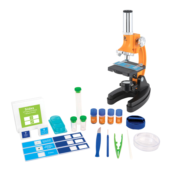
Discovery Telecom 900x Manual de instruções - Página 3
Procurar online ou descarregar pdf Manual de instruções para Microscópio Discovery Telecom 900x. Discovery Telecom 900x 5 páginas. Biological microscope

How do I use my microscope?
Before you use your microscope, make sure that the table, desk, or surface
that you place it on is stable and is not subject to vibration. If the microscope
needs to be moved, hold it by the arm and base while carefully transferring
it. Once the microscope is in a suitable location and the batteries are
installed, check the light source to make sure that it illuminates. Use a
microfiber cleaning cloth to gently wipe the lenses off. If the stage is dirty
with dust or oil, carefully clean it off. Make sure that you only raise and lower
the stage using the focus adjustment knob.
How do I operate the illumination?
Locate the mirror/light on the base of the microscope. Flip the mirror/light
to the "on" position (with the light facing up) and the light will illuminate.
This microscope is equipped with an incandescent light that illuminates the
specimen from below. The color filter wheel is located in the middle of the
microscope stage. The filters help you when you are observing very bright
or clear specimens. Using these filters, you can choose various brightness
levels and colors. This helps you better recognize the components of
colorless or transparent objects (e.g. sea salt).
How do I adjust my microscope correctly?
Place the microscope in a suitable location as described above, and sit in a
comfortable viewing position. Always start each observation with the lowest
magnification. Adjust the distance of the microscope stage so that the
stage is in the lowest position, farthest away from the turret head. Turn the
objective turret until it clicks into place at the lowest magnification (Objective
5x/100x). Note: Before you change the objective setting, always make sure
the microscope stage is farthest away from turret by rotating the focus knob.
Separating the stage and turret by rotating the focus knob will avoid causing
damage to the specimen slide or microscope. When starting an observation,
always start with the 5x/100x objective in the rotating head.
Did you know?
The highest magnification is
not always the best for every
specimen!
Magnification Guide
Eyepiece
Objective
Power
20x
5x
100x
20x
20x
400x
20x
45x
900x
How do I observe the specimen?
Sitting in your location with
adequate illumination chosen from
the color filter wheel, the following
basic rules should be observed:
Start with a simple observation
at the lowest magnification.
Position the object or specimen
in the middle of the stage under
the stage clips, centered over the
lower light. Focus the image by
rotating the focus knob until a
clear image appears
in the eyepiece.
Place the prepared slide directly under the objective on the microscope stage,
and secure it with the stage clips. The prepared slide should be located directly
over the lower illumination. Look through the eyepiece, and carefully turn the
focus knob until the image appears clear and sharp. Now you can select a
higher magnification by rotating to the 20x/400x objective turret. Higher levels of
magnification can be achieved by turning the objective turret to a higher setting
(45x/900x). Following this procedure creates a steady increase of magnification
without overpowering the view of the object. The following magnifications should
be considered: 100x, 400x, then 900x. Each time the magnification changes (due
to the objective change), the image sharpness must be readjusted with the focus
knob. When doing this, be careful because if you move the microscope stage too
quickly, the objective and the slide could come into contact and cause damage to
the slide or microscope.
For transparent objects (e.g. sea salt), light is projected by the lower light
traveling from below the stage, through the objective and eyepiece, and finally
into your eye. This process of light transmission is known as microscopy. Many
micro-organisms found in water, plant components, and the smallest animal
parts are transparent in nature. Opaque specimens, on the other hand, will need
to be prepared for viewing. Opaque specimens can be made transparent by a
process of treatment and penetration with the correct materials (media), or by
slicing. You can read more about creating specimens in the following Microscope
Experiments booklet.
Troubleshooting Table
Problem
Solution
No recognizable image
Turn on light
Readjust focus
Start with the
lowest power objective (5x)
No image
Center object on slide under
lowest power objective (5x)
No light
Replace batteries
Check on/o position
