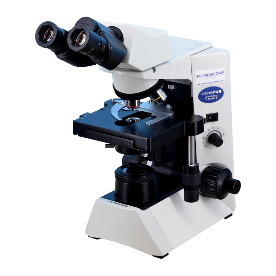
Olympus CX31 Руководство по эксплуатации - Страница 16
Просмотреть онлайн или скачать pdf Руководство по эксплуатации для Микроскоп Olympus CX31. Olympus CX31 24 страницы. Biological microscope

4-5 Condenser
Fig. 21
Fig. 22
Fig. 23
13
1
Centering the Field Iris Diaphragm
1. With the 10X objective engaged and the specimen brought into focus,
turn the field iris diaphragm ring @ counterclockwise to stop down the
diaphragm to near its minimum size.
2. Turn the condenser height adjustment knob ² to bring the field iris
diaphragm image into focus.
3. Rotate the two condenser centering knobs ³ to adjust so that the field
iris diaphragm image is centered in the eyepiece field of view. (Figs. 21
& 22)
4. To check centration, open the field iris diaphragm until its image
touches the perimeter of the field of view. If the image is not precisely
inscribed in the field of view, center again. (Fig. 22)
5. When used for actual observation, open the field iris diaphragm until its
image is slightly larger than the field of view.
2
Aperture Iris Diaphragm
· The aperture iris diaphragm determines the numerical aperture of the
illumination system. Matching the numerical aperture @ of the
illumination system with that of the objective provides better image
resolution and contrast, and also increases the depth of focus.
Adjustment method
Align the aperture iris diaphragm knob ² with the NA value @ on the
scale. The scale value should correspond to the NA number engraved
on the objective.
Since the aperture iris diaphragm knob has a certain width, align the
center line engraved on the knob with the scale indication.
When using a 100X objective, turn the aperture iris diaphragm knob ²
all the way to 0.9 on the scale. (Fig. 23)
· Since the contrast of microscope specimens is ordinarily low, setting
the condenser aperture iris diaphragm to between 70% and 80% of
the N.A. of the objective in use is usually recommended.
#If the aperture iris diaphragm is set too small, image ghost may be
observed.
(Figs. 21 & 22)
(Fig. 23)
