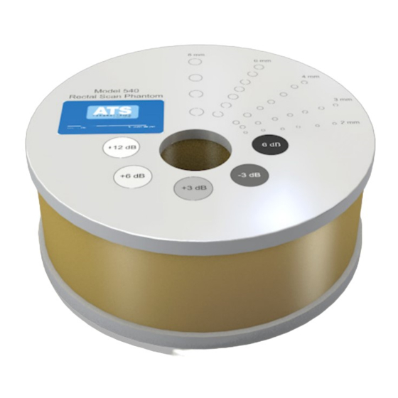
ATS 540 Series Kullanım Kılavuzu - Sayfa 7
Test Ekipmanları ATS 540 Series için çevrimiçi göz atın veya pdf Kullanım Kılavuzu indirin. ATS 540 Series 9 sayfaları. Rectal scan phantom

5. Move the transducer slightly until the anechoic circular targets are clearly imaged.
6. Freeze image and obtain a hard copy.
7. Examine the image to determine the first and last target in each size group displayed.
Record the range of depths visualized for each group. Due to the configuration of the
sound beam small targets in the near field may not be imaged.
8. All findings should be documented on the quality assurance record.
Results
The targets should appear circular with sharp clearly defined edges, indicating an
abrupt transition from the echogenic to the anechoic region. The targets are anechoic
and should be free of any internal echoes or fill-in. However, the presence of internal
system noise may manifest itself by producing an observable shade of gray within the
target area.
The specific values determined, while significant in their own right, are somewhat less
important than stability over time. Performing this test on a routine basis at the same
instrument settings should produce the same results. Any changes should be
investigated.
GRAY SCALE & DISPLAYED DYNAMIC RANGE
Description and Reason For Testing
Gray scale or gray scale processing uses the amplitude of the echoes received to vary
the degree of brightness of the displayed image. The adjustment of the echo signal
required to go from a just noticeable (lowest gray scale level) echo to the maximum
echo brightness is referred to as the displayed dynamic range. Clinically, gray scale
processing and displayed dynamic range allow echoes of varying degrees of amplitude
to be displayed in the same image.
Test Procedure
1. Place the phantom on a clean, flat surface with the internal scanning well positioned
for use.
2. Fill the scanning well slowly with water to avoid introducing air bubbles.
3. Insert the transducer into the scanning well.
4. Adjust the instrument settings (TGC, output, etc.) to establish baseline values for
"normal" liver scanning. If the bottom of the phantom is seen, adjust the gain settings
until the image fades and goes entirely black at the bottom of the display. Record these
settings on the quality assurance record. These setting should be used for subsequent
testing.
