Olympus IX70 Instructions d'utilisation - Page 2
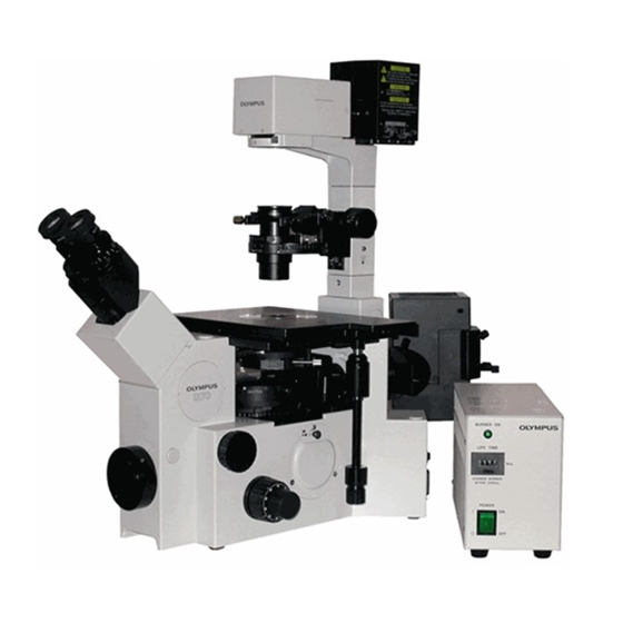
Parcourez en ligne ou téléchargez le pdf Instructions d'utilisation pour {nom_de_la_catégorie} Olympus IX70. Olympus IX70 5 pages. Inverted fluorescent microscope
Également pour Olympus IX70 : Manuel d'utilisation (32 pages)

Fluorescence Imaging Instructions:
1. Turn off transmitted light.
2. Rotate the filter wheel to your desired filter for your specific sample. (Green for
CY3, TRITC; blue of Alexa488 , FITC; UV for DAPI, etc.) (See Fig. 2 for filter
wheel)
a. You should see a colored light coming through the objective lens (i.e. blue,
green)
3. Find an area of your specimen that you want to image using the XY stage control
and fine focus knob.
Imaging Instructions:
1. Pull out slider to block the light to the objective lens (right side level with the
oculars)
a. Third stop, camera icon
2. Open the software on the computer: cellSens Entry
3. Click the live button to turn on the camera
4. Adjust the exposure by moving the slider to right until your image appears
properly exposed.
a.
The exposure normally can be set as auto in bright field image mode and as
manual in fluorescent image mode.
5. Normalize background:
a. For Bright field images: on the top right toolbar, click the white dropper icon
b. For fluorescent images: on the top right toolbar, click the black dropper icon
c. Then select an area of 'background ' on your image slide by drawing a small
box encompassing only the background. This sets the write or black balance for
your image and reduces background noise.
6.
Focus your image to the camera using the fine focus if needed.
7.
Scale Bar:
a. Select scale bar by going to view drop down on the top menu and click on
scal bar tab.
b. Make sure you have the correct value for the object lens
8. Click the Snapshot icon to capture image (next to the live button)
a. Save your image under the user folder on the desktop (if you have scale bar
on your images. When you save images as .tiff file, the images will have two
layers; when you save your images as .jpeg file, the images will only have one
layer)
9. When finished, close the software
10. Turn off microscope and mercury lamp.
··
