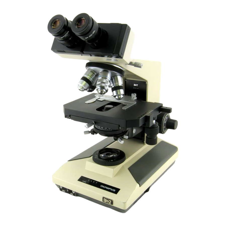
Olympus BH2 Series Manual - Page 3
Browse online or download pdf Manual for Microscope Olympus BH2 Series. Olympus BH2 Series 6 pages. Phase contrast
Also for Olympus BH2 Series: Troubleshooting & Adjustment Manual (46 pages), Complete Teardown, Cleaning, And Reassembly (23 pages), Complete Teardown, Cleaning, And Reassembly (40 pages), Teardown, Cleaning And Reassembly (45 pages), Installation Manual (9 pages)

Introduction
This document describes the proper procedures to set
up an Olympus BH-2 microscope for phase contrast
viewing.
Scope of this Document
The procedure described here was performed using an
Olympus BHT microscope, but this procedure also
applies to the BHS, BHSP, BHSU, BHTP, and BHTU
models as well.
What is Phase Contrast?
The technique of phase contrast microscopy was
developed in the 1930s by Dutch physicist Frits Zernike
and began to be broadly used by 1942. Zernike was
awarded the Nobel Prize in Physics for his achievement
in 1953. Phase contrast techniques are most useful for
studying living, non-stained specimens, since live
specimens cannot typically be stained without affecting
their behavior or killing them outright. For these types
of unstained specimens, phase contrast provides
significantly increased contrast as compared to
conventional brightfield microscopy.
phase contrast optics exaggerate the differences in the
phase relationships between the light waves in the
background illumination and the light waves passing
through the specimen, so that they can constructively
or de-constructively interfere with each other at the
intermediate image plane, thereby converting invisible
phase differences into visible image contrast.
How Does Phase Contrast Work?
In conventional brightfield microscopy, a visible image is
formed by wave interference at the intermediate image
plane of the background illumination (the background
illumination is the light that does not pass through the
specimen, which is also known as the "S", or surround
wave-front) and of the diffracted light (the diffracted
light is the light that passes through the specimen under
observation, which is also known as the "D", or
diffracted wave-front). The image of the specimen is
visible to the observer due to the diffraction,
absorption, and phase-shifting that occurs as the D
wave-front passes through the specimen under
observation. Diffraction in the D wave-front occurs as a
result of detail in the specimen. Absorption occurs as a
result
of the
specimen being not
transparent.
Phase shift occurs as a result of
differences in the refractive index of the specimen, as
compared to the surrounding medium.
When viewing live, unstained specimens in brightfield,
the specimen is often difficult to see since light
absorption
can
be
constructive/destructive interference that occurs as a
Phase Contrast on the Olympus BH-2 Microscopes
In a nutshell,
completely
minimal
and
since
result of the phase-shifted D wave-front is minimal as
well. Phase contrast microscopy utilizes special optics
(both in the condenser and in the objectives) to
accomplish two things: 1) The S wave-front (i.e., the
background illumination) is decreased in amplitude by
the phase ring in the objective so that the intensity of
the D wave-front will not be swamped by the otherwise
bright background lighting. 2) The S wave-front is phase
shifted by a quarter wavelength by the phase ring in the
objective, thereby exaggerating the constructive or
destructive interference that occurs between the S and
D wave-fronts at the intermediate image plane. The
result of these two things is that images of live,
unstained specimens have significantly higher contrast
than could otherwise be obtained using convention
brightfield microscopy.
Phase Contrast Types
Olympus phase contrast optics for the BH-2 are
available in two basic types: Positive and Negative, as
indicated by the markings on the objective barrel.
Positive phase contrast objectives are marked with a
"P" (such as "PL" or "PLL") and negative objectives are
marked with an "N" (such as "NH" or "NM").
In positive phase contrast, the phase ring in the
objective advances the S wave-front by a quarter
wavelength, relative to the D wave-front, whereas in
negative phase contrast, the phase ring retards the S
wave-front by a quarter wavelength, relative to the D
wave-front. In both cases, the phase of the D wave-
front is retarded by areas of the specimen which have a
higher refractive index than the surrounding medium,
and advanced by the areas of the specimen which have
a lower refractive index than the surrounding medium.
In positive phase contrast, the advanced S wave and the
retarded D wave destructively interfere at the
intermediate image plane, resulting in the areas of the
specimen with a higher refractive index than the
surrounding medium appearing darker than the neutral-
gray background. In negative phase contrast, the exact
opposite occurs. The retarded S wave and the retarded
D wave constructively interfere at the intermediate
image plane, resulting in the areas of the specimen with
a higher refractive index than the surrounding medium
appearing lighter than the neutral-gray background.
Setting Up for Phase Contrast Microscopy
The following equipment will be needed to utilize phase
contrast on Olympus BH-2 microscopes.
• Olympus BH-2 microscope
• BH2-PC or BH2-PCD Zernike-style phase contrast condenser
• Olympus CT-5 or CT-30 phase-centering telescope (as
the
appropriate)
Revision 1
Page 3 of 6
